
Many people picture the immune system as an army of white blood cells that hunts germs. The real picture is bigger. To understand how the immune system works, you need to see it as a defense network. This network joins physical barriers, chemical alarms, inflammation, precise antigen recognition, and long-term memory.
The immune system does not work like a conveyor belt. Its two arms, innate immunity and adaptive immunity, talk to each other all the time. Together they recognize threats, communicate, remove invaders, and regulate the response so it does not harm your own tissues.
In this post, you will learn each part of this network and how the parts work together.
1. The Starting Point: Pathogens vs. Host
The process starts when a pathogen tries to enter your body. A pathogen can be a bacterium, virus, fungus, or parasite. It can also be a product made by these organisms.
The biggest challenge is recognition. Your immune system must tell dangerous microbes apart from your own healthy cells. It must also ignore harmless substances from the environment.
To meet this challenge, your body builds its defense in layers:
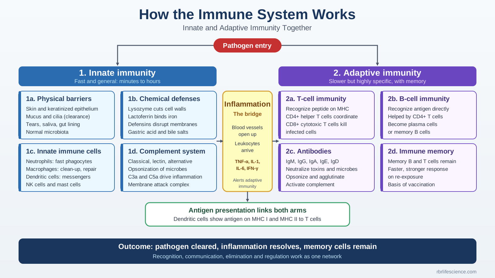
- Innate immunity: This layer acts within minutes to hours. It uses physical barriers, chemical defenses, complement proteins, and phagocytes. These parts recognize common microbial patterns and signs of tissue damage.
- Inflammation: This layer links the two arms. Inflammatory mediators change blood vessels, call white blood cells to the infection site, and help contain the threat. They also connect innate sensing to adaptive activation.
- Adaptive immunity: This layer usually responds more slowly the first time. However, it is highly specific. B and T lymphocytes use special receptors to target particular antigens. They also build immunological memory, so your body responds faster and stronger next time.
Innate and adaptive responses often happen at the same time and influence each other.
Want to learn more about antigens? Read our post on immunogens vs antigens.
2. Innate vs. Adaptive Immunity at a Glance
This table compares the two arms.
| Feature | Innate immunity | Adaptive immunity |
|---|---|---|
| Recognition | Common pathogen patterns and damage signals | Specific antigens, recognized by B-cell and T-cell receptors |
| Initial response | Rapid | Usually slower during the first exposure |
| Main components | Epithelia, phagocytes, NK cells, complement, and soluble mediators | B cells, T cells, antibodies, and memory cells |
| Receptor diversity | Receptors encoded in the germline | Highly diverse receptors made by gene rearrangement |
| Memory | No classical antigen-specific memory, although “trained immunity” can occur | Classical antigen-specific memory |
| Major role | Early containment and activation of other defenses | Targeted elimination and better protection on re-exposure |
Many people think innate immunity is “non-specific.” That is not fully correct. Innate receptors do recognize specific molecular patterns. They simply lack the huge diversity of lymphocyte receptors.
3. Innate Immunity: The First Line of Defense
Innate immunity has four main parts. Together, they stop invaders early.
Physical Barriers: Stopping Entry
Your first line of defense is not a passive wall. It is an active system.
- Skin: Keratinized epithelium forms a physical barrier. Surface lipids and acidic conditions add more protection.
- Mucus: Sticky mucus traps microbes and particles in your airways, gut, and other mucosal surfaces.
- Mucociliary clearance: Tiny hair-like cilia move mucus and trapped debris toward your throat, where your body clears it.
- Tears and saliva: They wash away microbes and contain antimicrobial substances.
- Gastrointestinal tract: Stomach acid, digestive enzymes, mucus, tight epithelial junctions, and antimicrobial peptides all help defend it.
- Normal microbiota: Helpful microbes compete with pathogens for food and attachment sites. They can also make substances that inhibit pathogens.
Epithelial cells do more than block entry. They sense microbes, release cytokines and chemokines, make antimicrobial molecules, and signal the immune cells beneath them. In your respiratory tract, for example, mucus traps inhaled particles and cilia sweep them out. If microbes cross the barrier, resident immune cells respond.
To explore where immune cells develop, see our post on primary lymphoid organs.
Biochemical Defense Mechanisms
Your body also uses chemicals to limit microbial growth. Saliva, sweat, tears, mucus, stomach acid, and bile all play a role.
| Defense factor | How it works |
|---|---|
| Lysozyme | Breaks bonds in bacterial cell walls (peptidoglycan) |
| Gastric acid | Creates a low-pH environment that damages or stops many swallowed microbes |
| Lactoferrin | Binds iron, so microbes cannot use it |
| Defensins | Disrupt microbial membranes and support epithelial defense |
| Bile salts | Disrupt some microbial membranes and reduce microbial survival |
| Mucus and fluid secretions | Trap, dilute, and help remove microbes |
Different cells make these factors, and not all of them are strictly “immune” products. For example, specialized stomach cells make gastric acid, not white blood cells.
General Immune Cells: The Cellular Arm
When a microbe breaches the barriers, innate immune cells step in. Each cell type has its own job.
- Neutrophils: These cells reach infection sites quickly. They engulf bacteria and fungi and kill them with toxic granules and reactive oxygen species. Read more in our post on neutrophils, the fast-acting immune cells.
- Macrophages: They engulf pathogens and cell debris, release warning cytokines, take part in inflammation, and help repair tissue.
- Dendritic cells: These cells act as messengers. They detect microbial signals, capture antigens, and show peptide fragments to T cells. In this way, they link innate and adaptive immunity.
- Natural killer (NK) cells: They patrol for virus-infected cells and tumor cells. They kill these cells without earlier antigen-specific sensitization. They also release cytokines such as interferon-gamma.
- Mast cells, eosinophils, and basophils: These cells drive allergic inflammation and defend against helminths (parasitic worms). Their exact roles depend on the cell type and the tissue.
These cells are not interchangeable, even though their jobs overlap. Neutrophils respond fast, macrophages defend and maintain tissue, dendritic cells connect to adaptive immunity, and NK cells kill infected cells.
The Complement System
Complement is a group of proteins in your blood and on cell membranes. These proteins activate each other in a cascade. Three pathways can start the cascade:
- Classical pathway: Antibodies bound to antigens usually trigger it. Some other recognition molecules can also trigger it. See our guide to the classical pathway of complement activation.
- Lectin pathway: Lectins start it when they recognize microbial carbohydrates.
- Alternative pathway: It begins with spontaneous C3 activation, which then amplifies on suitable surfaces, including many microbial surfaces. Read our notes on the alternative pathway.
All three pathways meet at C3 activation. They then achieve three main goals:
- Opsonization: C3b and related fragments coat microbes, so phagocytes can recognize and swallow them more easily.
- Inflammation: C3a and C5a send inflammatory signals. C5a also attracts and activates several types of leukocytes.
- Direct membrane attack: The membrane attack complex (C5b–C9) punches holes in some microbial membranes, especially those of certain Gram-negative bacteria.
Complement shows how well the immune system integrates its parts. It belongs mainly to innate immunity, yet antibodies from adaptive immunity activate its classical pathway. Complement is not a cell. It is a protein system that works beside the cells.
For a full overview, read our post on the complement system: pathways, functions, and regulation.
4. The Inflammatory Response: The Central Bridge
Acute inflammation links the fast innate response to the start of the adaptive response. It contains an infection where it begins, and it sends the signals that alert the adaptive immune system.
Inflammation starts when resident tissue cells, such as macrophages and mast cells, detect microbial components or tissue injury through pattern-recognition receptors. These cells release cytokines, chemokines, and lipid mediators. Mast cells also release histamine. Complement activation adds fragments such as C3a and C5a.
The Inflammatory Sequence
- Recognition: Pattern-recognition receptors on resident macrophages, mast cells, and dendritic cells detect microbial structures or damage signals.
- Mediator release: These cells release cytokines such as TNF-α, IL-1, and IL-6, along with chemokines and lipid mediators. Complement fragments add to the signal.
- Vascular changes: The mediators make blood vessels dilate and become more permeable. As a result, plasma proteins and white blood cells can leave the bloodstream and enter the tissue.
- Leukocyte recruitment: Chemokines and adhesion molecules on the vessel lining guide leukocytes into the tissue. In many acute bacterial infections, neutrophils arrive first.
- Elimination and resolution: Immune cells remove microbes and damaged material. If the response stays controlled, active resolution and tissue repair follow.
Learn more about complement’s role in this process in our post on complement in inflammation and regulation.
Key Cytokines and What They Do
People often group TNF-α, IL-1, IL-6, and IFN-γ together, but different cells release them, and each plays a distinct role:
- TNF-α and IL-1: Macrophages release them early. They activate the lining of blood vessels (the endothelium) and contribute to fever and body-wide inflammation.
- IL-6: It drives the acute-phase response and fever, and it influences B-cell responses.
- IFN-γ: NK cells and T cells produce most of it, so it usually appears later. It activates macrophages and supports cell-mediated immunity, especially against intracellular pathogens.
The exact mix of cytokines depends on the pathogen, the tissue, and the cells involved. Excessive or prolonged inflammation can damage healthy tissue, so the body must regulate it tightly.
Why Inflammation Is the Bridge
Inflammation does more than contain a local infection. The same microbial signals and inflammatory cytokines activate dendritic cells in the tissue. These dendritic cells mature, migrate to the lymph nodes, and present antigens, together with co-stimulatory signals, to naive T cells. This step starts the adaptive immune response.
Inflammation is not strictly required for every adaptive response. Still, in most infections it is the main coordination mechanism. Inflammation alone does not guarantee an adaptive response. The immune system also needs antigen presentation, co-stimulation, and proper lymphocyte activation.
5. Adaptive Immunity: Precision and Memory
Innate immunity gives fast, broad protection. Adaptive immunity gives highly specific protection and long-term memory. It has two branches: T-cell-mediated immunity and humoral immunity. They work differently, but they cooperate closely.
T-Cell-Mediated Immunity (Cellular Immunity)
T cells handle threats inside your cells, such as viruses. They also coordinate immune responses and destroy infected or abnormal cells.
How a T-cell response unfolds:
- Antigen presentation: Dendritic cells process antigens and show peptide fragments on MHC molecules.
- T-cell recognition: A T-cell receptor recognizes a specific peptide–MHC complex.
- Activation: Antigen recognition, co-stimulatory signals, and cytokines together decide whether and how a naive T cell activates.
- Clonal expansion: Activated T cells multiply. This creates many cells that recognize the same antigen.
- Differentiation: T cells develop into subsets that fit the threat.
- Effector activity: Effector T cells coordinate immune functions or carry them out directly.
The two major T-cell groups:
- CD4+ helper T cells: They recognize peptides on MHC class II. Depending on their subtype, they activate macrophages, help B cells make strong antibodies, recruit other immune cells, or regulate inflammation.
- CD8+ cytotoxic T cells: They usually recognize peptides on MHC class I. They kill virus-infected or cancerous cells with perforin and granzymes, or through death-receptor pathways.
T cells do not recognize free-floating germs the way B cells do. They rely on MHC (major histocompatibility complex) molecules. MHC class I shows internal proteins to CD8+ cells. MHC class II shows processed outside proteins to CD4+ helper cells.
Humoral Immunity (Antibody-Mediated Immunity)
B cells and antibodies drive humoral immunity. It neutralizes pathogens and toxins outside cells, blocks microbes from attaching, and helps remove them.
B-cell activation, step by step:
- A B cell recognizes an antigen through its B-cell receptor.
- In many protein-antigen responses, the B cell takes in the antigen and shows peptides to helper T cells.
- Helper signals, including CD40–CD40L interaction and cytokines, drive B-cell growth and differentiation.
- Some activated B cells become plasma cells, which are antibody factories.
- Other B cells become memory B cells, which support faster future responses.
Some antigens, such as certain repeating non-protein structures, activate B cells through T-independent pathways. Therefore, not every antibody response needs helper T cells.
What B cells can do: B cells recognize antigens with membrane-bound immunoglobulin. They show peptides to helper T cells, release cytokines, become plasma cells, and form memory B cells. Naive B cells are not dormant until an antigen shows up. They circulate constantly and scan lymphoid tissues.
What antibodies actually do:
- Neutralization: Antibodies bind pathogens or toxins and stop them from attaching to cells or causing harm.
- Opsonization: Antibodies coat microbes, so phagocytes recognize them through Fc receptors and take them up more easily.
- Complement activation: Some antigen-bound antibodies trigger the classical complement pathway.
- Agglutination: Antibodies cross-link particles or microbes and clump them together for easier removal.
The Five Antibody Classes
| Antibody | Main function |
|---|---|
| IgM | Usually the first class made in a primary response; strong at agglutination and complement activation |
| IgG | The main antibody in blood and tissues; supports neutralization, opsonization, and complement activation, depending on the subclass |
| IgA | Protects mucosal surfaces; common in saliva, tears, and breast milk |
| IgE | Drives immediate allergic reactions and defends against some parasites |
| IgD | Works mainly as a receptor on mature naive B cells, together with IgM |
The heavy-chain class sets an antibody’s effector functions. Its antigen-binding region decides which antigen it recognizes.
6. The Missing Link: Antigen Presentation and MHC
Antigen presentation turns a fast, general innate response into a specific, targeted adaptive defense. The innate system does not simply hand a pathogen over to adaptive immunity. It actively shapes how the adaptive system responds.
How Antigen Presentation Links Both Arms of Immunity
- Capture in peripheral tissues: Dendritic cells belong to the innate cellular arm. They patrol tissues, meet pathogens, and take them in, often by phagocytosis.
- Processing and migration: The dendritic cell breaks microbial proteins into small peptide fragments. It then matures and travels through the lymphatic vessels to the lymph nodes, where naive T cells wait.
- MHC display: In the lymph node, the dendritic cell shows these peptides on its surface using MHC molecules.
- MHC class II presents peptides to CD4+ helper T cells, which coordinate the wider immune response.
- MHC class I presents peptides to CD8+ cytotoxic T cells, which kill infected or abnormal cells. MHC class I normally shows proteins made inside the cell. Dendritic cells can also load peptides from outside antigens onto MHC class I through cross-presentation, which lets them activate CD8+ T cells.
- Co-stimulation and cytokine signals: Presentation alone is not enough. Dendritic cells also provide co-stimulatory signals and cytokines. Together, these signals decide whether a naive T cell activates and which subtype it becomes.
| Feature | MHC class I | MHC class II |
|---|---|---|
| Where it appears | Most nucleated cells | Mainly professional antigen-presenting cells (dendritic cells, macrophages, B cells) |
| Typical antigen source | Proteins made inside the cell | Proteins processed through endosomal pathways |
| Recognized by | CD8+ T cells | CD4+ T cells |
| Main role | Lets cytotoxic T cells spot infected or abnormal cells | Lets helper T cells coordinate immune responses |
Why This Link Matters
T cells cannot recognize free, intact antigens the way B cells can. Their receptors only recognize peptides displayed on MHC molecules. Dendritic cells capture threats through innate mechanisms and turn them into MHC–peptide complexes. In this way, they help the adaptive immune system mount a precise and well-matched response.
How the Immune System Works Together: A Viral Infection Example
A respiratory viral infection shows the whole network in action.
- Entry and barrier defense: The virus meets mucus, epithelial cells, and mucociliary clearance. If it reaches susceptible cells, infection can begin.
- Innate recognition: Infected epithelial cells and resident immune cells detect viral parts through pattern-recognition receptors. Interferons and other mediators set up local antiviral defenses.
- Inflammation and recruitment: Cytokines and chemokines bring in reinforcements. The strength of this response depends on the virus and the host.
- Antigen presentation: Dendritic cells collect viral pieces, travel to lymph nodes, and show them to T cells with the right co-stimulation.
- Cell-mediated response: CD8+ T cells kill infected cells. CD4+ T cells coordinate the wider response.
- Humoral response: B cells become plasma cells and release neutralizing antibodies. These antibodies reduce infection of new cells.
- Resolution and memory: The body clears the virus, inflammation settles, and tissue repairs. Memory B and T cells stay behind to protect you in the future.
These steps overlap. Innate defenses keep working while adaptive immunity develops. In turn, adaptive effectors strengthen innate mechanisms. Not every infection follows the same timeline or uses every mechanism equally.
Conclusion
Now you know how the immune system works. Its parts never act alone. Barriers, innate recognition, inflammation, complement, antigen presentation, lymphocyte responses, and memory all depend on each other. Innate immunity buys time and sets the stage. Adaptive immunity then delivers a precise and lasting defense. Regulatory mechanisms keep the whole system from harming your own tissues.
For a broader view, see our immune system overview of organs and cell lineages.