
[lwptoc titleFontWeight=”extrabold” itemsFontSize=”90%” colorScheme=”dark” backgroundColor=”#d1fff9″ borderColor=”#28f0ff” titleColor=”#000000″ linkColor=”#2167d1″ hoverLinkColor=”” visitedLinkColor=”#1e73be”]
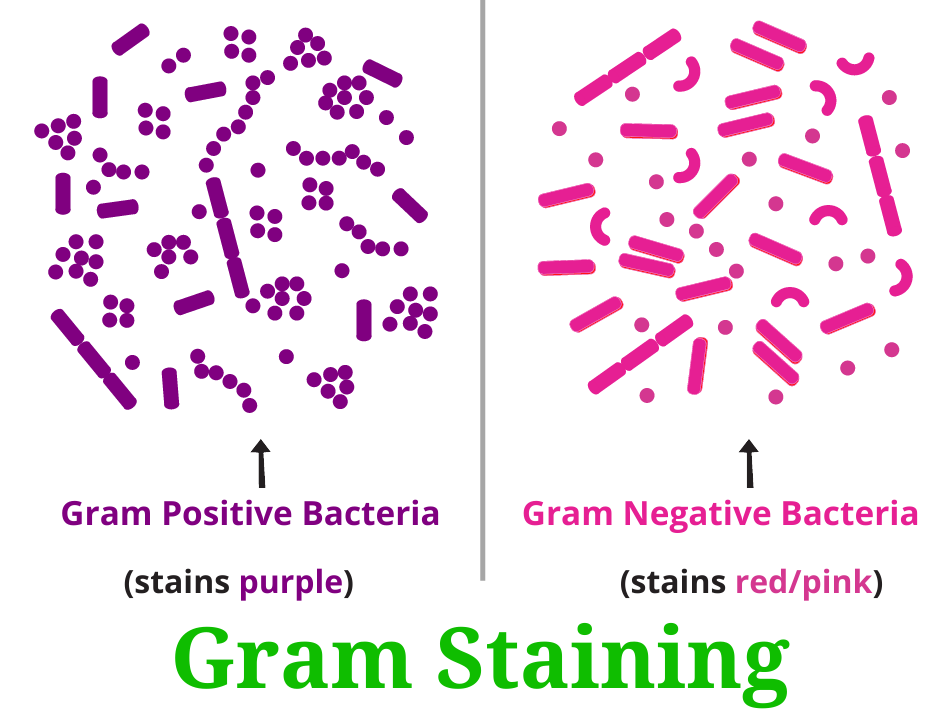
Gram staining procedure is a popular and widely used method in microbiology. To study and identify any new bacteria usually the first step performed is gram staining. In 1884 Danish Physician Dr. Hans Christian Gram developed this staining procedure. Gram’s staining is the differential staining procedure. Gram staining divides bacteria into two categories as gram-positive bacteria and gram-negative bacteria. After successful gram staining, gram-positive bacteria appear purple and gram-negative bacteria will appear reddish-pink.
Principal of Gram staining
In the gram staining procedure bacteria are first stained with the primary stain crystal violet. Crystal violet is fixed using mordant Iodine. There is a physical and chemical difference in the composition of the cell wall of the bacteria. Some types of bacteria have a complex and thick peptidoglycan layer in their cell wall. Whereas some types of bacteria have a thin peptidoglycan layer in their cell wall.
The thick and complex peptidoglycan layer in the cell wall causes the primary stain Crystal Violet to get trapped in the cell wall. This crystal violet doesn’t leach out upon treatment with alcohol. Bacteria retaining the primary dye crystal violet even after the decolorization step appear as purple are known as gram-positive bacteria.
Bacteria with thin peptidoglycan layer in their cell wall, the primary stain Crystal Violet doesn’t get trapped in the cell wall. The crystal violet leaches out easily upon treatment with alcohol and bacteria becomes colorless. Such bacteria who lose the primary dye complex (crystal violet) after the decolorization step and uptake, the counterstain safranin, and appear as reddish-pink are known as gram-negative bacteria.
Reagents used for gram staining include
- Crystal Violet- Primary stain
- Grams Iodine solution- The mordant which fixes crystal violet to cell wall
- Alcohol (95%) or Acetone – The decolorizer
- Safranin- The counterstain
Steps in gram staining
There are two steps in staining. First step is preparation of smear on a glass slide. Second step is gram staining this smear with grams reagents.
Step 1 : Preparation of smear on a glass slide
- Take a loopful of bacterial culture sample on a clean and grease-free slide.
- Make a smear on the slide, Air dry, and heat fix the smear. (Gently pass the slide through the flame 2-3 times)
Step 2 : Gram Staining
- Add Crystal Violet on smear and stand for 1 min
- Gently Wash the slide using tap water
- Add gram’s Iodine solution for 1 min
- Gently Wash the slide using tap water
- Add Ethyl alcohol (95 %) stand for 15 -30 seconds
- Gently Wash the slide using tap water
- Add counterstain safranin stand for 1 min
- Observe the slide under the microscope with a 100 X objective oil immersion lens.
Watch below the nice video on gram staining procedure
Role of Gram reagents used for gram staining:
Primary stain: Crystal violet
Crystal violet is the primary stain in grams staining procedure It is a basic stain it stains both gram-positive and gram-negative bacteria in purple color.
Mordant: Grams Iodine solution
The mordant in the gram staining is the Grams Iodine. Iodine helps to fix the dye crystal violet in the cell wall Iodine reacts with crystal violet and forms an insoluble Crystal violet –Iodine complex (CV-I). Due to this complex color of the stain become intense. After the application of mordant both gram-positive and gram-negative bacteria appear in purple color.
Decolorizer : Ethanol 95 % or Acetone
In gram staining ethanol (95 %) or Acetone works as a decolorizer. The Decolorizations step is the most critical step in the gram staining technique. Alcohol plays a dual role. Alcohol dehydrates the proteins and also dissolves the lipid. . In gram-positive bacteria peptidoglycan layer is thick. When Alcohol dehydrates the thick peptidoglycan layer the pores in the cell wall shrink and become small in size. The peptidoglycan layer becomes tight. Thus, CV-I complex is insoluble and not accessible by alcohol. Large primary stain CV-I complex is trapped in the peptidoglycan layer. Alcohol doesn’t wash out the CV-I complex from the cell wall of gram-positive bacteria. And gram-positive bacteria retain purple color in their cell wall after treatment with alcohol.
In gram-negative bacteria, the peptidoglycan layer is thin and surrounded by an outer membrane. Gram-negative bacteria have more lipid in their outer layer. Alcohol dissolves this lipid layer and the cell wall becomes porous. Alcohol washes out the primary stain CV-I complex from the thin and less complex peptidoglycan layer. So gram-negative cells become colorless after treatment with alcohol
Counterstain: Safranin
Safranin acts as the counterstain in grams staining.
In the final step, Safranin stains only colorless bacterial cells.
Safranine stains colorless gram negative cells to reddish- pink color.
Why do gram-positive bacteria stain Purple color in gram staining?
Gram-positive bacteria have a thick peptidoglycan layer in the cell wall. The primary stain Crystal violet is fixed in a thick peptidoglycan layer by the Mordant Grams iodine. Crystal Violet-Iodine complex is insoluble and remains trapped inside the thick peptidoglycan layer. In the Decolorization step alcohol doesn’t wash out this complex. In the next step counterstain, safranin doesn’t stain because the Crystal Violet-Iodine complex is already occupied the space in the peptidoglycan layer. So the cell shows the color of crystal violet i.e. Purple. That’s why gram-positive bacteria stain purple in grams staining.
Why do gram-negative bacteria stain pink color in gram staining?
The cell wall of gram-negative bacteria is complex. The thin peptidoglycan layer is surrounded by the additional membrane known as the outer membrane. This outer membrane contains more lipid. Most of the lipids get dissolved by alcohol during decolourization step. Alcohol easily washes out Crystal Violet-Iodine complex from thinner peptidoglycan layer. Such cells appear colorless. In next step counterstain safranin stain such colorless cells to pink-red color. That’s why gram-negative bacteria stain pink-red in grams staining.
Gram-positive bacteria: Examples
Staphylococcus aureus, Streptococcus, Streptococcus pneumoniae, Clostridium diphtheria, Clostridium tetani, Bacillus anthracis, Clostridium botulinum, Clostridium perfringens, Streptococcus pyogenes
Gram-negative bacteria: Examples
Escherichia Coli, Haemophilus influenzae, Salmonella typhi , Shigella spp, Vibrio cholerae, Neisseria gonorrhoeae ,Neisseria meningitidis, Pseudomonas aeruginosa, Yersinia pestis
The difference in cell wall composition makes bacterial cells react differently to gram stain.
Solve MCQ on Gram Staining Procedure
Click here to solve MCQSimilar posts like this Acid-fast staining method
Thanks,RBR Life sciences
Good
Thank you very nice explan
Good day sir I am Ismail Ibrahim from kaiji Niger state bargu local government please sir here is my complain about all laboratory practical I want on everything sir thank for concen my problem good luck
Thanks RBL 😊
Thanks for you sir 😊😊
Good
Sir,that was really helpful for me thank you sir
Very nice explanation sir thankyou for your suitable description.
Thanks rbrlifescience! Huge shoutout to Ma’am Basas, proud student here <3 !