
Microorganisms such as bacteria are very small, colorless, and transparent. As a result we cannot see these microorganisms with naked eyes. The reason why we can’t see microorganisms is that they lack contrast. In other words, without contrast and color, to see such transparent microbial cells is difficult. To observe and identify any microbes, firstly, we need to stain them with dyes. Dyes create the contrast between microbial specimens and their background. In short, staining with dyes enhances visualization of microbes under a light microscope. Depending upon the number of dyes used and purpose, staining is categorized into different types.
Three different types of staining in microbiology:
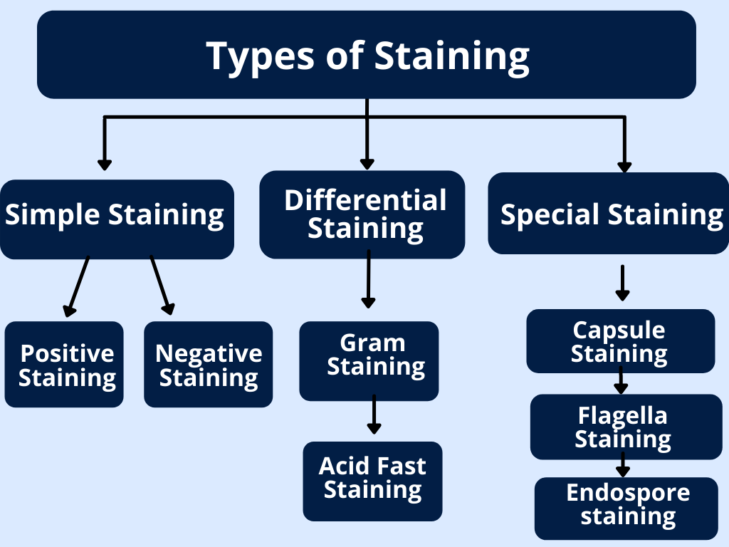
In microbiology, there are three main types of staining techniques . These are simple staining, differential staining, and special staining (see flowchart ). The purpose of use of all these three staining techniques is different. For example, A simple staining technique visualizes the morphology, size, and shape of microorganisms. Likewise, differential staining distinguishes various types of microbial and bacterial groups. Similarly, special staining demonstrates structures of bacteria such as flagella, capsules, and endospores.
1) Simple staining:
The simple staining method uses a single dye to stain bacterial specimens. It is easy and very simple to use. This staining requires basic dyes. Bacterial cell surfaces have negative charge so basic dyes easily bind to cell surface having negative charge. Most importantly, simple staining is useful to study the size, shape, and arrangement of bacterial cells. There are two types of simple staining procedure.
a) Positive staining:
Positive staining is direct staining. This method uses basic dyes. Firstly, the bacterial smear is stained with single basic dyes and bacterial cells take up the stain. After that, cells appear colored but, the background is white.
b) Negative Staining:
Negative Staining method is Indirect staining. Here the background takes up the stain. This method uses acidic dyes. Bacterial cells don’t take up the stain. Bacterial cells look transparent against the dark background.
2) Differential Staining:
IIn this procedure we use more than one dye for staining. Different types of bacteria have variations in structural properties. So different types of bacteria will react differently to differential stains. Therefore two types of bacteria stains in two different colors. A differential staining procedure distinguishes different groups of bacteria. For examples Gram staining and Acid Fast staining are differential staining procedures.
a) Gram staining:
Gram staining procedure divides bacteria into two groups. These are gram-positive bacteria and gram-negative bacteria.
b) Acid-fast staining:
Acid-fast staining differentiates bacteria into two groups. These are Acid-fast bacteria and non-acid fast bacteria.
3) Special staining:
Bacteria have specific internal and external structures. These structures include flagella, capsule layer, and endospores. These structures vary between different bacterial groups. A Special staining procedure has importance in bacterial identification and taxonomy. This staining procedure allows you to observe such specific structures. Special staining procedure require complex stains. There is use of more than one dye for this staining. Below are the examples of special staining procedures in microbiology.
a) Capsule staining:
Many bacteria contain a special layer surrounding their cell wall. This layer is capsule. The capsule staining is done in order to observe the capsule layer.
b) Flagella staining:
Some bacteria have Flagella. It helps bacteria in movement and motility. If you do flagella staining then you can observe the presence and arrangement of flagella on bacteria.
c) Endospore staining:
Endospore is a dormant structure of bacteria. It forms during unfavorable survival conditions. With Endospore staining one can observe free spores and spore structure in bacteria.
To sum up, different types of staining procedure are important tools in microbiology. These staining methods are used for preliminary study of microbes.